The influence of nano-structural interfaces and their composition on bone’s fracture behavior

Human cortical bone is a highly complex composite that attains its unique combination of strength and toughness through deformation and toughening mechanisms at multiple length-scales throughout its hierarchical framework. Ageing and disease-related factors may alter the structure of bone at all levels of its hierarchy, raising the risk of fracture. A key aspect of maintaining bone’s mechanical properties is its ability to remodel and repair damaged tissue.
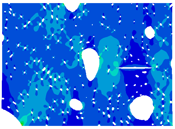
One main result of the remodeling process is the so-called cement line, which surrounds the newly formed cylindrical bone unit (i.e., secondary osteon) and serves as a highly mineralized and stiff interface between the remaining bone material and the remodeled bone material. Cement lines are thought to play a crucial role in the nanomechanical properties of bone: Growing cracks have been observed to be deflected at cement lines, thereby increasing bone toughness at the sub-microscale. Furthermore, ageing will cause that the distribution of the osteocyte lacunae as potential stress concentrators and/or crack barriers within the osteon gets changed and affects the strength and toughness of bone at small length scales, which consequently affects the global bone quality and fracture characteristics.
Here, we propose an in-depth study of the bone fracture properties in young and aged individuals by taking advantage of the unique nano imaging and crystallography modalities at the German Electron Synchrotron (DESY), image processing and analysis, and computational multi-scale modeling techniques available at the University Medical Center Hamburg-Eppendorf (UKE).


